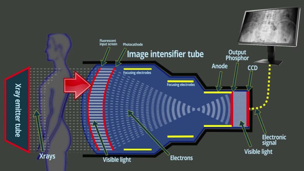
An image intensifier is a device used in medical imaging to amplify and convert low-intensity X-ray or fluoroscopic images into bright, high-contrast images that are visible on a display screen. It consists of a vacuum tube with a photocathode at one end and a phosphor screen at the other end, connected by a series of electron optics. Here are some key points about image intensifiers:

-
Operation: When X-rays or fluoroscopic radiation pass through the patient’s body, they strike the input phosphor of the image intensifier, causing it to emit light photons. The photocathode, which is sensitive to light, converts these photons into electrons via the photoelectric effect.
-
Electron Acceleration: The electrons emitted by the photocathode are accelerated and focused by a series of electrodes within the vacuum tube. These electrodes create an electric field that accelerates the electrons towards the output phosphor screen.
-
Image Amplification: As the accelerated electrons strike the output phosphor, they cause it to emit a brighter and more intense light image than the original input phosphor image. This amplification process increases the brightness and contrast of the image by thousands of times, making it visible to the human eye or to an electronic detector.
-
Fluoroscopy: Image intensifiers are commonly used in fluoroscopy procedures, where real-time X-ray images are needed to visualize dynamic processes within the body, such as the movement of organs, blood flow, or the progress of medical procedures like catheter insertion or joint injections.
-
Radiography: Image intensifiers can also be used in radiography systems to enhance the visibility of low-intensity X-ray images, particularly in situations where high-contrast images are required, such as in vascular imaging, orthopedics, and interventional radiology procedures.
-
Components: The main components of an image intensifier include the input phosphor, photocathode, electron optics (such as focusing electrodes and accelerating electrodes), and output phosphor. Some modern image intensifiers may also incorporate digital imaging technology for image capture and processing.
-
Applications: Image intensifiers are widely used in various medical specialties, including radiology, cardiology, neurology, vascular surgery, and orthopedics, for both diagnostic and interventional procedures. They play a critical role in improving image quality, reducing radiation dose, and enhancing patient care.


